


Ongoing research is moving beyond the canonical Akt signaling pathway and transforming the understanding of the mechanisms that regulate glucose transport into the skeletal muscle, according to Paul M. Titchenell, PhD, University of Pennsylvania, who chaired the Friday, June 21, symposium The Second Act— Redefining the Signaling Mediators of Glucose Uptake in Muscle.
The session can be viewed on-demand by registered meeting participants on the virtual meeting platform. If you haven’t registered for the 84th Scientific Sessions, register today to access the valuable meeting content through Aug. 26.
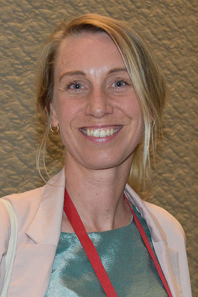
Lykke Sylow, PhD, University of Copenhagen, Denmark, said evidence for Akt as a major regulator of glucose uptake in skeletal muscle is limited. Dr. Sylow and colleagues have identified the Rho guanosine triphosphate (GTPase) Rac1 as an important mediator of the final step of mobilizing vesicles containing the glucose transporter GLUT4 to the plasma membrane. Rac1 activity is diminished in muscle cells from both individuals with obesity and those with type 2 diabetes, likely due to upregulation of the Rac1 inhibitor guanine dissociation inhibitor alpha (GDIɑ).
Given that skeletal muscle is a major sink for glucose, Dr. Sylow believes that targeting insulin resistance in these cells could be a novel therapeutic strategy in type 2 diabetes.
“Molecular pathways that mediate glucose uptake in skeletal muscle represent an untapped opportunity for the treatment of type 2 diabetes,” she said. “As a new regulator of insulin-stimulated glucose uptake, the Rac1-GDIɑ complex may represent a way to increase glucose in muscle and normalize glycemia.”
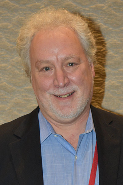
Jonathan Bogan, MD, Yale University, discussed an additional aspect of regulating GLUT4-containing vesicles to the plasma membrane. He showed that the protein TUG sequesters GLUT4-containing vesicles to the Golgi apparatus within the cell. In the presence of insulin, proteolytic cleavage of TUG releases the vesicles, he explained. They are then trafficked to the plasma membrane, where GLUT4 can facilitate the uptake of glucose into the cell. The resulting C-terminal domain of TUG is also released and can drive the expression of genes involved in oxidative metabolism and thermogenesis.
“GLUT4 storage vesicles represent a stable pool of vesicles near the endoplasmic reticulum and Golgi apparatus,” Dr. Bogan said. “In the setting of insulin resistance, this pool of vesicles is depleted, and we also see impaired TUG cleavage. This means the cell no longer has an insulin-responsive vesicle pool that can be mobilized to the cell surface in response to high blood glucose levels.”
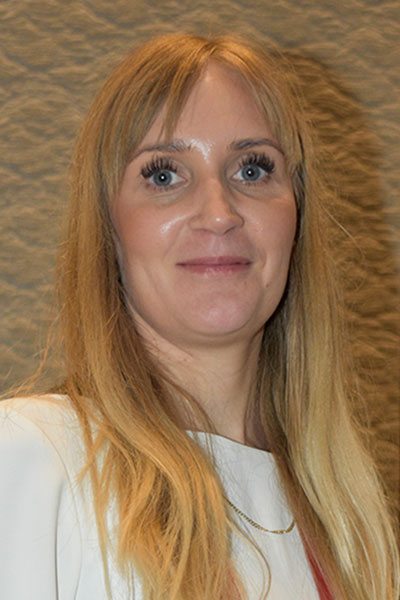
While Dr. Sylow and Dr. Bogan discussed new players to the canonical insulin pathway, Katrin Svensson, PhD, Stanford University, presented work on a new, insulin-independent pathway to increase glucose uptake. She showed that the protein isthmin-1, which is secreted by adipose cells, has some of the same activities as insulin. In skeletal muscle cells, isthmin-1 activates the Akt/PI3K pathway, resulting in increased glucose uptake via GLUT4. However, unlike insulin, isthmin-1 does not signal through the insulin receptor. It also has differential effects on lipid synthesis. While insulin induces lipogenesis and protein synthesis, isthmin-1 blocks lipogenesis, especially when insulin levels are high.

“Because isthmin-1 comes from adipose tissue, it increases in conditions of obesity as a way to counteract insulin resistance,” Dr. Svensson said. “Isthmin-1 targets about 50 percent of the same targets as insulin,” she continued, “but there are some targets unique to isthmin-1, and these change the way cells use energy.”
P. Darrell Neufer, PhD, East Carolina University, demonstrated how energy production in the mitochondria is linked to insulin sensitivity and glucose metabolism. As electrons flow through the electron transport chain to drive the synthesis of ATP, some inevitably leak out of the system, interacting with oxygen and hydrogen to form hydrogen peroxide (H2O2). In the setting of overnutrition, emission of H2O2 by the mitochondria increases, which ultimately changes the redox state of the cell. In mice, scavenging redox-active species, either pharmacologically or genetically, are protected against insulin resistance. Dr. Neufer proposed that there may be a redox relay that interferes with the transport of GLUT4 to the plasma membrane.
“Overnutrition puts a lot of reducing pressure on the mitochondria, generating a lot of H2O2,” he said. “Eventually the cell and the redox state of various proteins begins to shift. The failure to keep these proteins in a reduced state has been linked to diabetes and other diseases.”

Register Today for the 85th Scientific Sessions
Join us in Chicago for the 85th Scientific Sessions, June 20–23, to learn about the latest advances in diabetes research, prevention, and care. Full in-person registration includes access to all of the valuable onsite content during the meeting and on-demand access to session recordings June 25–August 25.